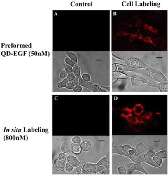
Figure 3.
Targeting of QDs to A431 (squamous cancer) cells using norbornene-tetrazine cycloaddition. Top: QD fluorescence at 605 nm with excitation at 488 nm. Bottom: corresponding DIC images (scale bar 10 μm). Cells were targeted either by (B) using preformed QD-EGF complexes (50 nM) for single QD tracking or by (D) performing in situ conjugation using norbornene-functionalized QDs (800 nM) on BATEGF- modified cell surfaces for ensemble labeling. (A) and (C) are control experiment with poly(PEG12)-PIL QDs (without norbornene).

