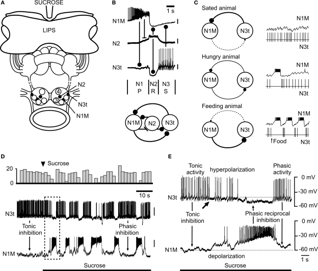
Figure 2.
Modulation of the snail feeding system by tonic inhibition (adapted from Staras et al., 2003, with permission from Elsevier). (A) The semi-intact preparation used for electrophysiological recording showing the location of feeding CPG interneurons in the buccal ganglia. A feeding stimulus, sucrose, is applied to the lips. (B) Firing patterns (top) and synaptic connections (bottom) of the three main types of interneurons that form the feeding central pattern generator. One cycle of feeding activity is shown with the origins of the synaptic inputs indicated by connecting bars. The N1M fires during the first protraction (P) phase, the N2 (with truncated spikes) the second rasp (R) phase and the N3t the third swallow (S) phase of the feeding cycle. (C) Model summarizing the modulatory effects of satiation and hunger on the tonic inhibition of the feeding pattern. In satiated animals the N3t fires continuously and the consequent inhibitory effects on the N1M prevent bursting in this cell. In hungry animals, even with no food present, there are occasional feeding bursts in the N1M due to the lower rate of firing of the N3t. In feeding animals the tonic N3t is weak and insufficient to prevent sustained bursting in the N1M and the N3t fires phasically to become part of the feeding rhythm. The thickness of the continuous lines connecting the N1M and N3t cells (left) indicates the strength of the inhibitory effects between the cells and the dashed lines the absence of phasic inhibitory effects. (D) An experiment in the semi-intact preparation showing that a food stimulus reduces the suppressive inhibitory control by the N3t cell and releases rhythmic fictive feeding activity. The change in the pattern of N3t firing is emphasized by the top trace where the number of N3t spikes is accumulated in 3s bins. (E) Expansion of the boxed area shown in (D) shows the first cycle of fictive feeding activity in the N1M after the sucrose-induced reduction in N3t firing rate. The arrow under the N3t trace indicates the point at which N3t starts to hyperpolarize and its tonic spike frequency begins to decrease. This decrease in N3t firing is followed by complete suppression of N3t firing when the N1M becomes active and synaptically inhibits the N3t. The subsequent phasic N1M-N3t reciprocal inhibition leads to the alternating patterns of N1M/N3t firing seen throughout the feeding pattern that follows. In (A) to (C) a stick indicates an excitatory synaptic connection between neurons and a ball indicates an inhibitory synaptic connection.

