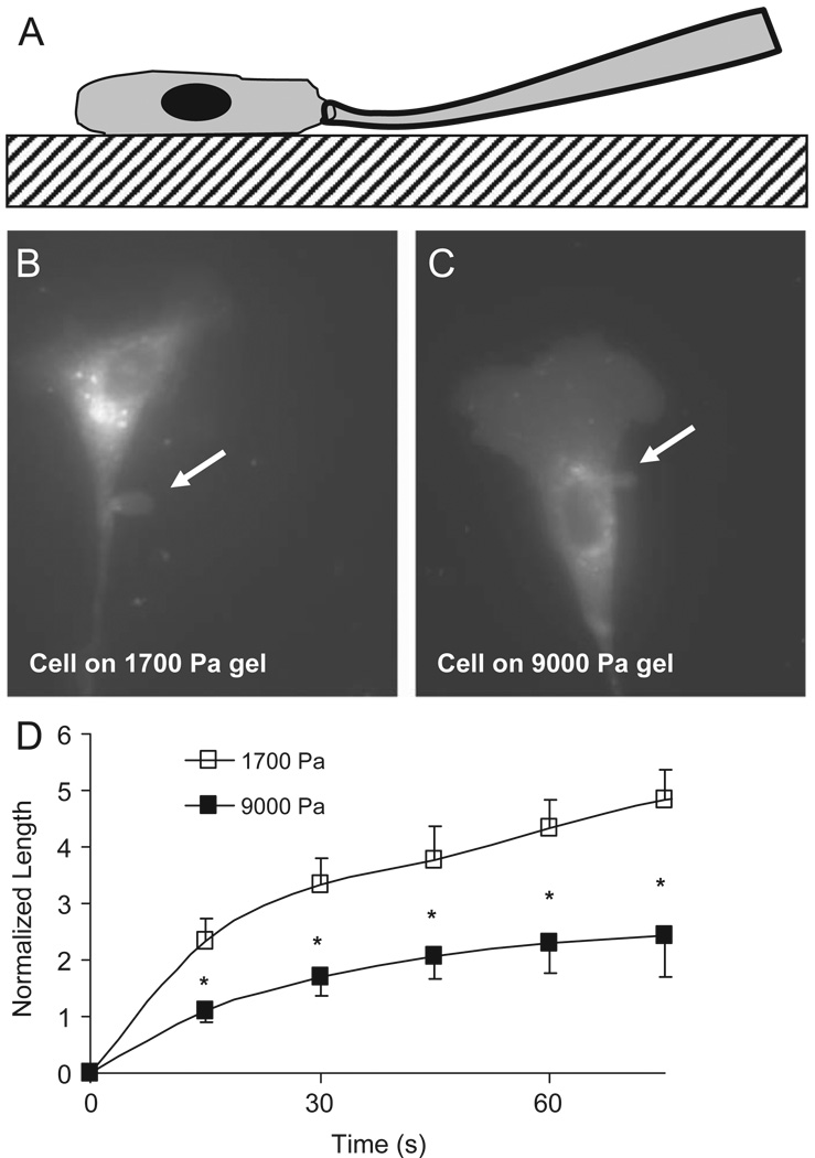
Fig. 1.
Microaspiration of adhered cells. The micropipette is gently pressed against the glass slide and slid into contact with the adhered cell (A). A vacuum is applied; aspirating a portion of the cell (arrow) into the pipette bore (B, C). The aspirated length is monitored as a function of time revealing that bovine aortic endothelial cells cultured on stiff substrates are more difficult to deform relative to those cultured on compliant gels (D).

