
Fig. 1.
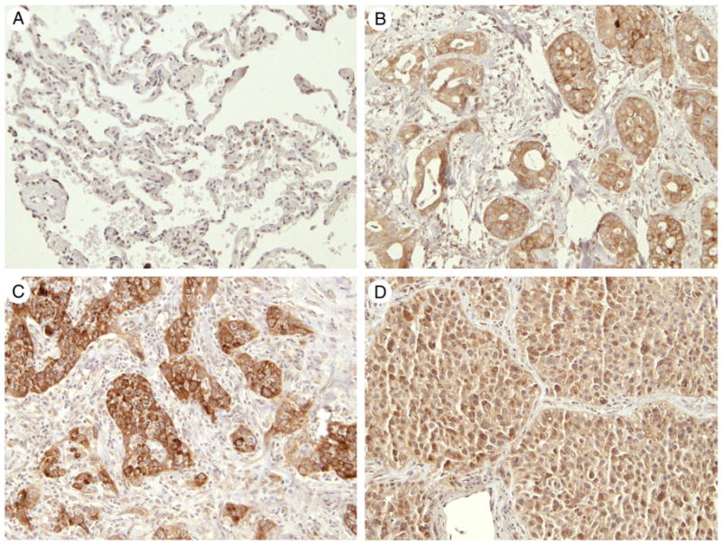
FB50 immunohistochemical staining in representative normal and cancerous lungs. (A) Normal lung showing no immunostaining of the alveolar and interstitial cells but weak staining of some alveolar macrophages, (B) positive immunostaining in adenocarcinoma tumor, (C) squamous cell carcinoma, and (D) large cell carcinoma (A-D, original magnification ×20).