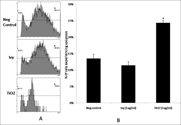
Figure 3.
Cytotoxicity analysis. HeLa cells were incubated with or without nanoparticles for 24 hours and stained with propidium iodide. The cell apoptosis was then determined by detection of fluorescence using flow cytometry. A) Representative flow cytometry plots for each of the three samples: negative control (Neg control), ivy nanoparticle (Ivy), TiO2 nanoparticle (TiO2). B) Cells experiencing apoptosis in the three samples. Each point represents an average of 3 samples from one of three experiments. * denotes significant difference based on Student's t test (p < 0.05).

