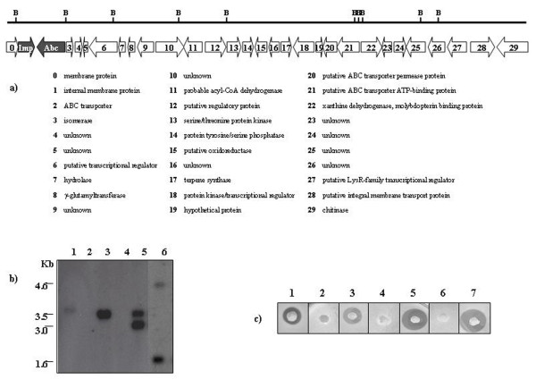
Figure 1.
Sequence analysis of the SL-40 insert and Bioassay. a) Schematic diagram of sequence analysis of clone SL-40. Putative ORFs are indicated by the arrows; below is a list of the protein which each ORF is most similar to. The genes involved in SL-3.5 antibacterial activity are shown in grey and the remaining ORFs are in white. B indicates BamHI sites. b) Southern blot analysis of BamHI-digested total DNA isolated from SL-3.5 (lane 1), S. lividans (lane 2), P. rosea (lane 3), SL-ESAC (lane 4), SL-Abc (lane 5) and SL-Imp (lane 6). The BamHI fragment of 3.5 Kb (derived from digestion of ESAC3.5) was used as probe. c) Bioassay against Micrococcus luteus: 1) SL-40; 2) SL-ESAC; 3) SL-3.5; 4) SL-Abc; 5) SL-Abc/Imp; 6) SL-Imp and 7) SL-Imp/Abc.

