Figure 1.
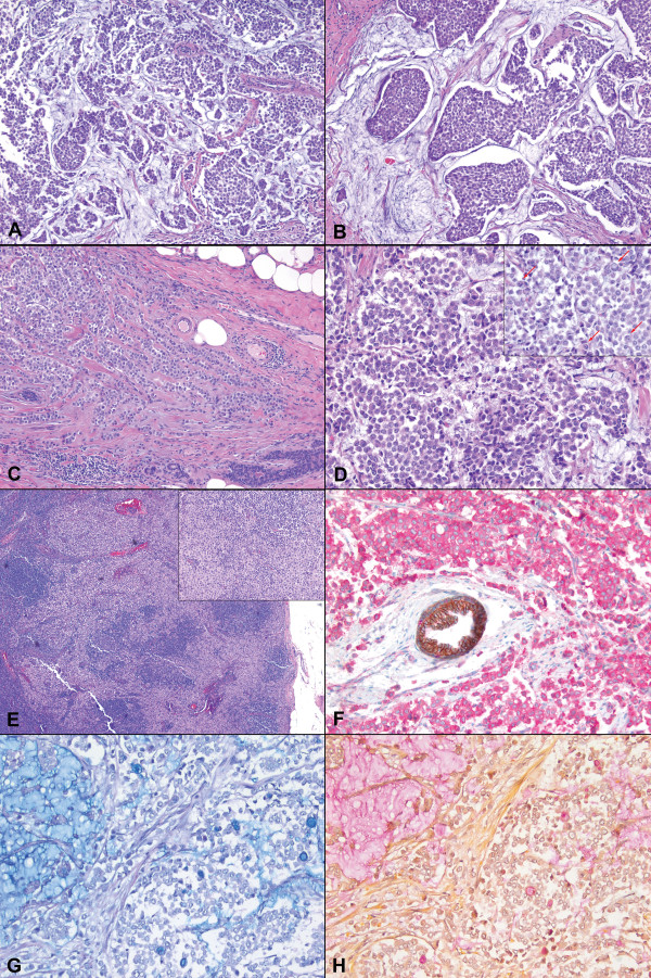
A case of invasive lobular carcinoma of breast with extracellular mucin. The tumor showed discohesive (A, H&E, ×100) and nested (B, ×200) growth with abundant extracellular mucin. At the periphery, the tumor cells formed cords and single files (C, ×100). Cytologically, the tumor cells were small to medium in size and relatively uniform (D, ×400), with scattered signet ring cells present (D inset, arrows, ×400). A sentinel lymph node showed metastatic focus of uniform tumor cells (E inset, ×40) in dissociated infiltrating pattern (E, ×200). Immunohistochemical stain revealed complete absence of membranous E-cadherin staining (brown) and the presence of diffuse cytoplasmic p120 staining (pink); a normal duct with membranous staining for both E-cadherin and p120 served as an internal control (F, immunohistochemistry, ×400). Alcian Blue (G, ×200) and Mucicarmine (H, ×200) stains highlighted both extracellular and intracellular mucin.

