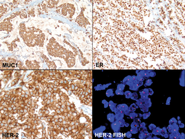
Figure 2.
Characteristics of the invasive lobular carcinoma with extracellular mucin. The tumor cells revealed strong cytoplasmic MUC1 staining (top left, immunohistochemistry, ×200), but were negative for MUC2, MUC4, MUC5AC and MUC6 (images not shown). The tumor cells were positive for ER (top right, ×400), negative for PR (image not shown), and positive for HER-2/neu with 3+ staining (bottom left, ×400). FISH study confirmed the amplification of HER-2 gene (bottom right, ×1,000).

