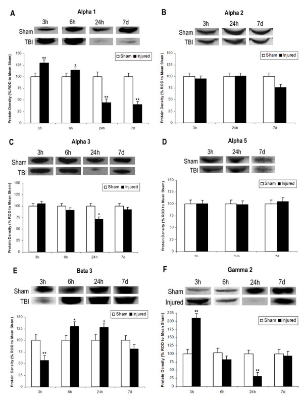
Figure 1.

Expression of GABAAR Subunits After TBI. Western blot analysis of GABA-A receptor subunits α1, α2, α3, α5, β3, and γ2 in the hippocampus 3 h, 6 h, 24 h, or 7 days after TBI. Histograms of protein expression were measured in Relative Optical Density (ROD) proportions normalized against the mean sham OD for each individual blot. Asterisks indicate significant differences based on factorial ANOVA; *p < .05, **p < .01. Error bars represent +/-SEM. A: Alpha 1 demonstrated significantly increased expression 3 h and 6 h post-TBI followed by significantly decreased expression at 24 h and 7 days. B: There were no significant differences in Alpha 2. C: Alpha 3 demonstrated significantly decreased expression at 24 h post-TBI only. D: There were no significant differences in Alpha 5. E: Beta 3 demonstrated initially significant decreased expression at 3 h, followed by significantly increased expression at 6 h and 24 h post injury. F: Gamma 2 demonstrated significantly increased expression at 3 h and significantly decreased expression at 24 h post-TBI.
