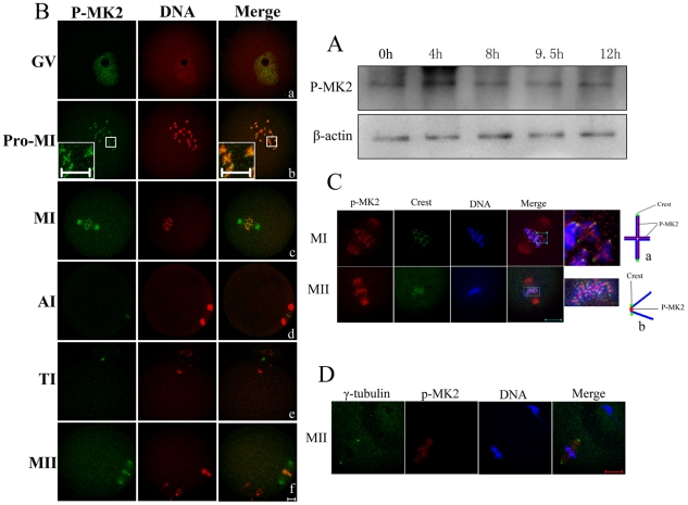
Figure 1. Expression and subcellular localization of p-MK2 during mouse oocyte meiotic maturation.
Samples were collected after oocytes had been cultured for 0, 4, 8, 9.5 and 12 h, corresponding to GV, Pro-MI, MI, AI to TI and MII stages, respectively. Proteins from a total of 200 oocytes were loaded for each sample. (B) Oocytes at various stages were double stained with antibodies against p-MK2 (green) and with propidium iodide (PI, red). GV, oocytes at germinal vesicle stage; Pro-MI, oocytes at first prometaphase; MI, oocytes at first metaphase; AI, oocytes at anaphase; TI, oocytes at telophase; MII, oocytes at second metaphase. Bar 20 µm. (C) Metaphase I and metaphase II oocytes were fixed and double labeled with rabbit p-MK2 antibody (red) and human Crest antibody (green). Each sample was counterstained with Hoechst 33258 to visualize DNA (blue). Bar 20 µm. (a) model chart for localization of p-MK2, Crest and chromosomes at MI stage oocytes. (b) model chart for localization of p-MK2, Crest and chromosomes at MII stage oocytes. (D) Oocytes cultured for 12h (MII) were fixed and stained for γ- tubulin (green), p-MK2 (red) and DNA (blue) as visualized with Hoechst 33258 staining. Bar 20 µm.

