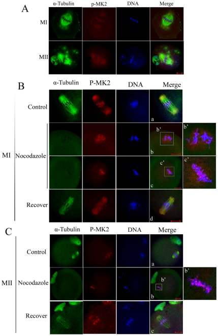
Figure 2. Localization of p-MK2 in mouse oocytes treated with taxol and nocodazole.
(A) Oocytes cultured for 8.5 h (MI) and 12 h (MII) were incubated in M16 medium containing 10 µM taxol for 45 min and then double stained with antibodies against p-MK2 (red), α-tubulin (green), and DNA (blue). (B) Oocytes at the metaphase I stage were incubated in M16 medium containing 20 µg/ml nocodazole for 10 min (b,c) and then washed thoroughly and cultured in fresh M16 mediun for 30 min (d). Control groups were treated with DMSO (a). Then oocytes were fixed and double stained with antibodies against p-MK2 (red), α-tubulin (green), and stained for DNA (blue). Bar 20 µm. (C) Oocytes at the metaphase II stage were incubated in M16 medium containing 20 µg/ml nocodazole for 10 min (b) and then washed thoroughly and cultured in fresh M16 medium for 30 min (c). Control group was treated with DMSO(a). Then oocytes were fixed and double stained with antibodies against p-MK2 (red), α-tubulin (green), and DNA (blue). Bar 20 µm.

