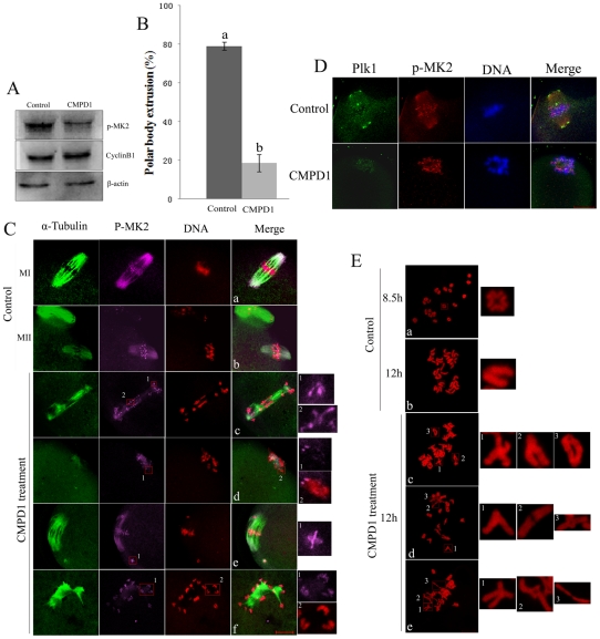
Figure 4. MK2 inhibitor CMPD1 treatment impairs spindle organization and chromosome alignment in mouse oocytes.
(A) Samples from control and CMPD1 groups were collected to test the efficiency of the MK2 inhibitor. CMPD1, 200 oocytes were cultured in M16 medium with 30 µM CMPD1 for 10 h; Control, 200 oocytes were cultured in M16 medium with DMSO for 10h. (B) The rate of oocytes with first polar body in the control group (n = 267) and CMPD1-treated group (n = 318). Data are presented as mean percentage (mean ± SEM) of at least three independent experiments. Different superscripts indicate statistical difference (p<0.05). (C) Spindle morphologies and chromosome alignment in oocytes cultured with DMSO or MK2 inhibitor CMPD1. GV oocytes were cultured in M16 medium with DMSO or with 30 µM CMPD1 for 12 h and then stained for p-MK2 (purple), α-tubulin (green) and DNA (red). Bar 20 µm. (D) GV oocytes were cultured in M16 medium with DMSO or with 30 µM CMPD1 for 8.5 h and then stained for p-MK2 (red), Plk1 (green) and DNA (blue). Bar 20 µm. (E) Chromosome spreading was performed in oocytes that had been cultured for 8.5 h or 12 h of DMSO treatment (MI) (MII) or for 12 h of CMPD1 treatment. Representative images of each sample are shown. Bar 10 µm.

