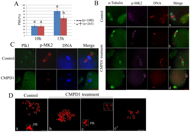
Figure 5. CMPD1 treatment leads to metaphase II spindle assembly defects, misaligned chromosomes and aneuploidy.
(A) The rate of oocytes with first polar body in the control group and CMPD1-treated group. Control group (−−): oocytes cultured with DMSO for 15 h (n = 100); CMPD1-treated group (−+): oocytes cultured with DMSO for 10 h, and then with 30 µM CMPD1 for 5 h (n = 265). Data are presented as mean percentage (mean ± SEM) of at least three independent experiments. Different superscripts indicate statistical difference (p<0.05). (B) Spindle morphologies and chromosome alignment in the oocytes first cultured with DMSO for 10 h, and then with MK2 inhibitor CMPD1 for 5 h; then stained for p-MK2 (purple), α-tubulin (green) and DNA (red). Control group oocytes were cultured with DMSO for 15 h. Scale bar, 20 µm. (C) GV oocytes were treated as in (B), and then stained for p-MK2 (red), Plk1 (green) and DNA (blue). Bar 20 µm. (D) Chromosome spreading was performed in oocytes that had been treated as (B). Representative images for each sample are shown. Bar 10 µm.

