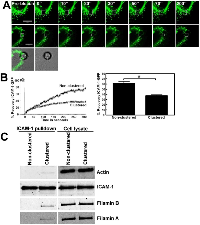
Figure 2. Lateral mobility of ICAM-1 depends on its C-terminal domain.
(A) ICAM-1-GFP is expressed in HeLa cells and FRAP was measured. Box in the upper panel depicts the FRAP region (square) and the recovery in time after every 10 seconds. Image at the right shows recovery after 200 seconds. Middle panel shows ICAM-1-GFP FRAP that is clustered around an adherent bead, coated with ICAM-1 antibodies. Lower image shows the merge of the green channel and DIC, in which the bead is visible. Bars, 10 µm. (B) Quantification of the FRAP data show decreased ICAM-1-GFP mobility following clustering. Graph on the left shows ICAM-1-GFP FRAP in time and graph on the right shows the difference in ICAM-1 mobility at t = 300 seconds. ICAM-1 was clustered using αICAM-1 antibody-coated beads. ICAM-1 FRAP was significantly reduced upon clustering. Experiment is carried out 6 times. Data are mean ± SEM. *p<0.01. (C) TNF-α-pretreated HUVECs were cultured on FN-coated surfaces and anti-ICAM-1-coated magnetic beads were allowed to adhere to the endothelium for 30 minutes, resulting in clustering of ICAM-1, after which the cells were lysed. As a control, the beads were added to endothelial cell lysates. Lysate conditions prevent ICAM-1 from clustering and these samples are therefore marked as Non-clustered. Next, using a magnetic pen, the beads were isolated from the cell lysates, washed and analyzed using Western blotting. The images on the left show that the magnetic beads efficiently precipitated ICAM-1 and that ICAM-1 clustering resulted in the recruitment of actin and filamin A and B. Images on the right show the expression of indicated proteins in the cell lysates.

