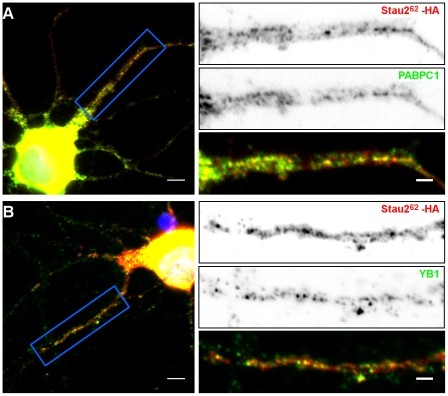
Figure 6. Co-localization of Stau262-HA3 with endogenous PABPC1 and YB1 in hippocampal neurons.
Neurons were transfected with a plasmid coding for Stau262-HA3. Twenty four hours post-transfection, neurons were fixed and labeled with anti-HA antibody (red) and either anti-YB1 (A) or anti-PABPC1 (B) antibodies (green). Left: Fluorescence microscopy of hippocampal neurons in culture. Scale bars: 5 µm. Right: Higher magnification of images showing protein localization in dendrites. The lower panels represent the superposition of both green and red signals. Scale bars: 2 µm.

