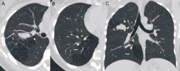
Figure 2.
Multi-detector computed tomography scan. (a) Axial image shows a mucus-like density opacity at the right upper lobe. The bronchial branch for the right upper lobe is not identifiable. (b) Axial image of the right lower lobe allows the characterization of a malformed multicystic area of the lung parenchyma. (c) Malformative features in minimum intensity projection (minIP) coronal reconstruction (bronchial atresia: thin arrow; congenital cystic adenomatoid malformation: thick arrow).

