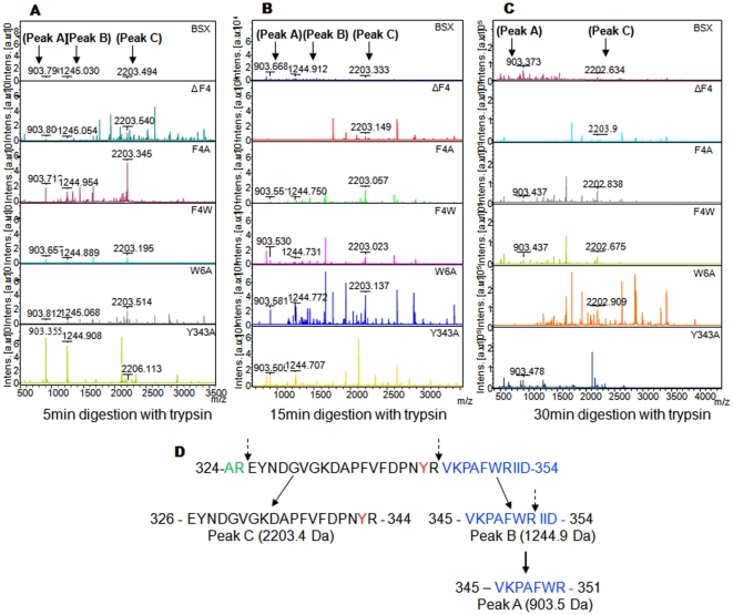
Figure 6. Limited proteolysis profile of BSX and its mutant.
All protein samples were incubated with trypsin for 5 min (A), 10 min (B) and 15 min (C) under native conditions and analyzed by mass spectrometry. (D) Schematic representation of limited proteolysis of R-BSX by trypsin toward the C-terminus. A partial C-terminal sequence of R-BSX from 326 to 354 is shown. Tyrosine 343 involved in the aromatic stacking interaction is shown in red. The dashed arrow indicates the trypsin cleavage sites.

