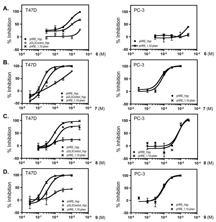
Figure 1. Concentration-response effects of 6 – 9 on HIF-1 activity in T47D (left panel of AD) and PC-3 (right panel of A – D) cell-based reporter assays.
Data shown are averages from one representative experiment performed in triplicate and the bars represent standard error. The label “pHRE_Hyp” indicates that the cells were transfected with the pHRE-TK-Luc reporter to monitor the ability of HIF-1 to direct the expression of luciferase under the control of hypoxia-response element (HRE) and the transfected cells were exposed to hypoxic conditions for 16 h. The label “pGL3Control_Hyp” indicates that the cells were transfected with a control plasmid pGL3-Control (Promega) that expresses luciferase under the control of the constitutively active CMV promoter and exposed to hypoxic conditions for 16 h. The label “pHRE_1,10-phen” indicates that cells transfected with the pHRE-TK-Luc reporter were treated with a hypoxia mimetic 1,10-phenanthroline (10 μM) for 16 h.

