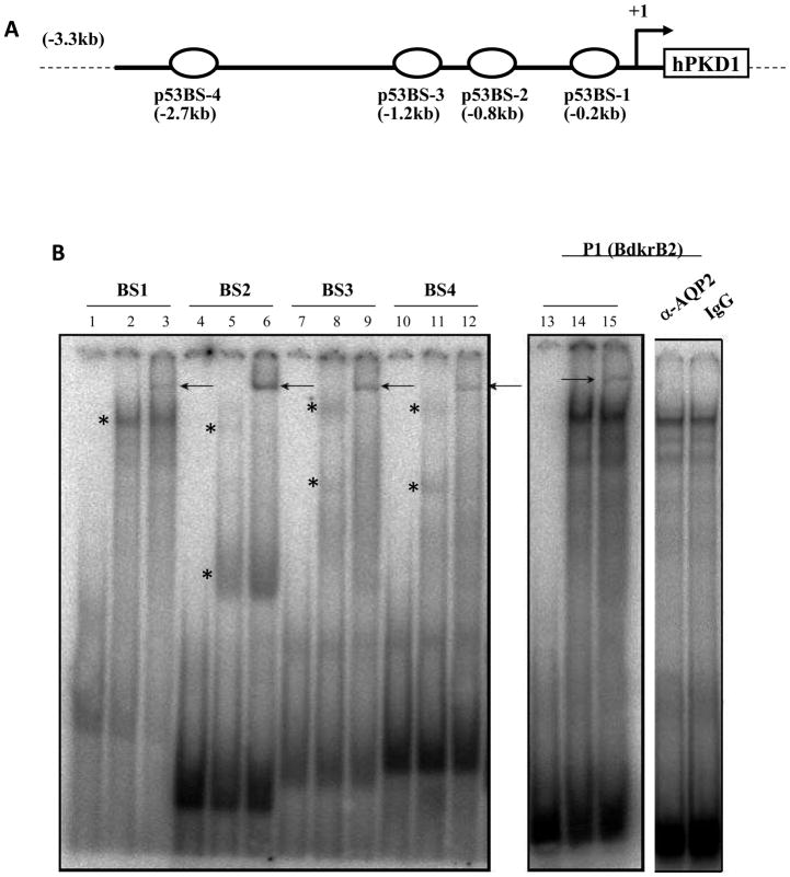
Figure 1. In vitro binding of p53 to the PKD1 promoter.
A. Schematic diagram showing the location of p53 consensus sequences, BS1-4. B. Electrophoretic mobility Shift Assays (EMSA). 32P-labeled oligoduplexes representing PKD1 BS1-4 were incubated with 3.5 μg of nuclear extract from IMCD3 cells. Lanes 1, 4, 7, 10 and 13: free probes; Lanes 2, 5, 8, 11 and 14: p53 BS1-4 and nuclear extract; Lanes 3, 6, 9, 12 and 15; BS 1-4 and nuclear extract in the presence of antibody against p53. Arrow indicates p53 specific super shift. P1 is an oligoduplex of the highly conserved p53 consensus sequence in the rat bradykinin B2 receptor promoter (positive control). Aquaporin 2 antibody and rabbit IgG served as negative controls.

