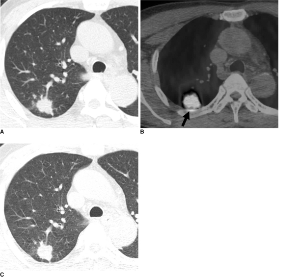
Fig. 1.
Pulmonary cryptococcosis with clustered nodular pattern in 53-year-old man who had hepatocellular carcinoma (patient 11 in Tables 1, 2).
A. Lung window of transverse CT scan obtained at level of distal trachea demonstrates clustered nodules in posterior segment of right upper lobe.
B. PET/CT scan obtained at similar level to A demonstrates high FDG uptake (arrow) within main nodule (maximum standardized uptake value, 9.3), hence simulating malignant nodule.
C. Follow-up CT scan obtained at similar level to and three months after A demonstrates slightly increased extent of clustered nodules in right upper lobe.

