Fig. 4.
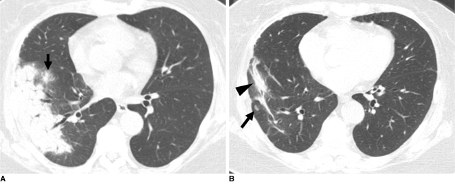
Cryptococcosis of bronchopneumonic pattern in 72-year-old woman who has underlying breast cancer (patient 22 in Tables 1, 2).
A. Lung window of transverse CT scan obtained at level of right inferior pulmonary vein shows subpleural consolidation and nodule (arrow) with surrounding halo in right lower lobe.
B. Follow-up CT scan obtained five months after A and with four months of anti-fungal therapy demonstrates remaining lesions of consolidation (arrowhead) and nodule (arrow) in right lower lobe.

