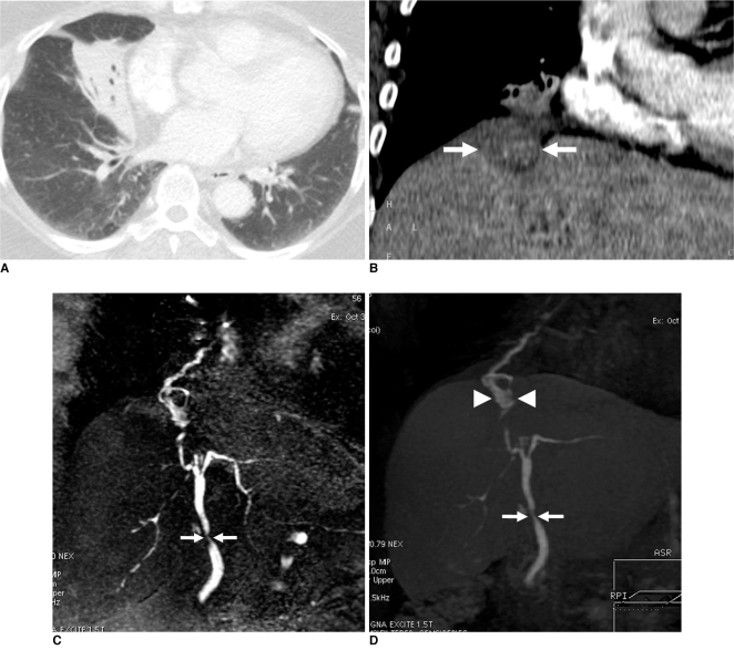
Fig. 1.
Bronchobiliary fistula in 56-year-old woman.
A. Chest CT shows distal subsegmental atelectasis and air bronchograms in right middle lobe.
B. Targeted coronal reconstruction of upper abdominal CT shows obscure subdiaphragmatic hypodense lesion (arrows) adjacent to inferior surface of right middle lobe.
C. Coronal 3D-maximum intensity projection reconstruction of conventional MR cholangiography demonstrates stenosis of common bile duct (arrows) as well as fistulous bronchobiliary communication.
D. Coronal 3D-maximum intensity projection reconstruction of contrast-enhanced MR cholangiography reveals contrast agent leaking from ventrocranial branch of right hepatic duct and further into subphrenic liver cyst (arrowheads), which transdiaphragmatically communicates with bronchial tree. Stricture of common bile duct is also demonstrated (arrows).

