
Figure 5.
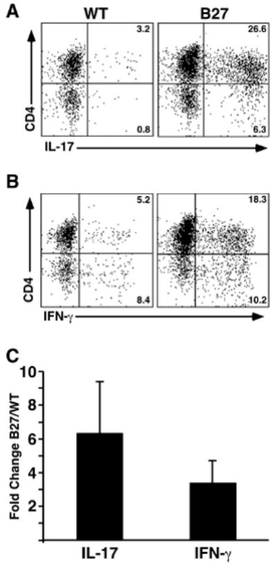
Expansion of CD4+ T cells expressing IL-17 in colonic lamina propria from HLA-B27 transgenic rats. Lamina propria lymphocytes were isolated from WT and HLA-B27 transgenic colon on a percoll gradient. Cells were analyzed for cell surface antigen and intracellular cytokines using FACS. Cells were gated on CD3+ and compared for cytokine production. A, Percentage of CD4+/IL-17+ and CD4-/IL-17+ cells in WT and HLA-B27 transgenic rats. B, Percentage of CD4+/IFN-γ+ and CD4-/IFN-γ+ cells in WT and HLA-B27 transgenic rats. C, Average fold-change in percentage of IL-17-expressing CD4+ T cells (IL-17; CD4+/IL-17+) and IFN-γ-expressing CD4+ T cells (IFN-γ; CD4+/IFN-γ+) populations between WT and HLA-B27 transgenic rats. Data shown are representative of three separate experiments using at least two animals per genotype and error bars represent standard error of the mean. Th17 cells are increased ∼6.3-fold (CI 3.1), and Th1 ∼3.4-fold (CI 1.3).