Fig. 1.

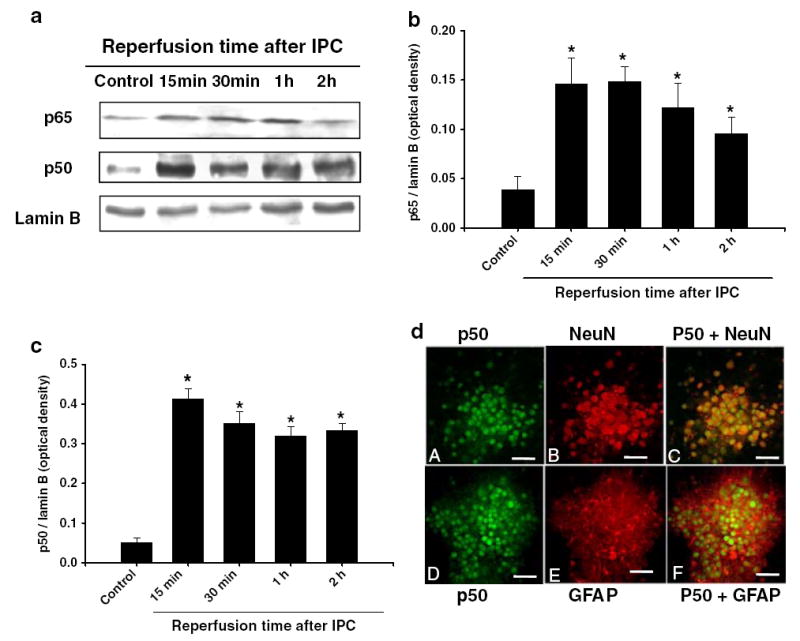
Ischemic preconditioning induced p65 and p50 translocation to the nucleus: a cells were lysed immediately after 1 h of IPC and at 15 min, 30 min, 1 h, and 2 h of reperfusion after 1 h of PC. Western blotting for p65 was carried out with nuclear protein extracts and the membrane was re-probed with the p50 antibody or nuclear lamin B antibody. b and c Histogram depicted densitometric analysis of Western blots of p65 or p50 in nuclear protein extracts compared with lamin B, respectively, *p<0.05 compared with control (n=5). d Confocal microscopic images of mixed cortical neuron/astrocyte cell cultures depicting co-localization of immunoreactivities for neuronal-specific antibody, NeuN (red) or astrocyte-specific antibody GFAP (red) and p50. At 30 min of reperfusion after IPC, NeuN (arrows; a, b, c) and GFAP (d, e, f) positive cells expressed p50. Bar:20μM
