
Fig. 6.
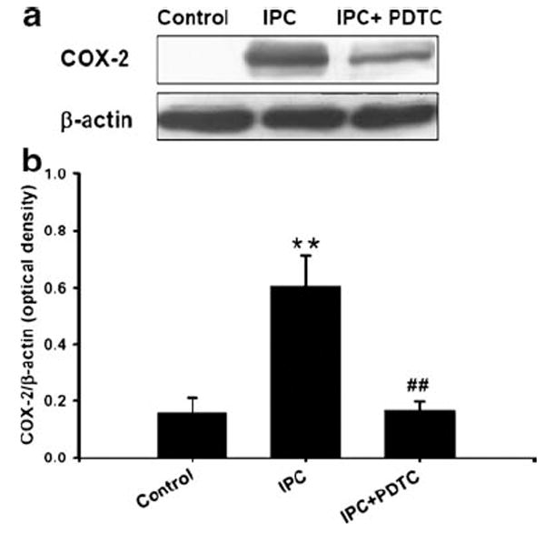
Inhibition of NF-kB reduced preconditioning-induced cyclooygenase-2 (COX-2) expression: a cells were treated with NF-kB inhibitor (PDTC, 1 μM) or vehicle during 24 h of reperfusion after 1 h of IPC. Cells were lysed at 24 h of reperfusion and analyzed by Western blotting with COX-2 antibody. The membrane was re-probed with β-actin antibody. Histogram depicted densitometric analysis of Western blots of COX-2 compared with β-actin. **p<0.01 compared with control, #p<0.05 compared with PC (n=3). b Histogram depicted densitometric analysis of Western blots of COX-2 and β-actin, *p<0.05 compared with control (n=5)