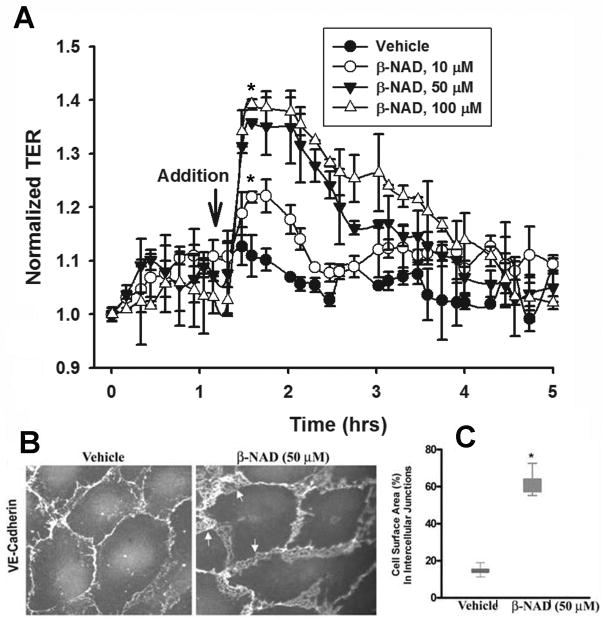
Figure 1.
Extracellular β-NAD enhances barrier function of HPAEC monolayers and increases VE-cadherin presentation in cell-cell contacts. (A) Dose-dependent TER response of β-NAD. HPAEC were challenged with increasing concentration of β-NAD (10–100 μM). Data are representative of several independent experiments (minimum of three) (*p < 0.05 compared with control). (B) Immunofluorescence staining of VE-cadherin in quiescent and β-NAD-stimulated HPAEC monolayers. The cells were treated with 50 μM β-NAD for 30 min, then fixed and immunostained using VE-cadherin antibody. Appreciably more VE-cadherin was recruited to the area of cell-cell junctions after β-NAD treatment. Arrows indicate overlapping edges of neighboring cells. (C) Quantification of the surface area of the cell-cell interface. The percentage of total cell-surface area occupied by VE-cadherin-positive cell-cell junctions was calculated for 20 cells in each group. The graph demonstrates that β-NAD induced a significant increase in cell-cell interface surface area as a percentage of total cell surface area (*p < 0.05 compared with control). The box and Whiskers plot show the means (lines at the box centers, 17.42% and 61.03% for control and β-NAD-treated cells respectively).

