Figure 5.

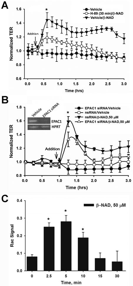
Molecular mechanisms of β-NAD-activated endothelial barrier enhancement in HPAEC. (A) β-NAD treatment activated cAMP-dependent PKA. HPAEC were pretreated with either vehicle or PKA-specific inhibitor, 5 μM H-89, for 30 min and then stimulated with 50 μM β-NAD in TER measurement assay. The inhibitor pretreatment significantly attenuated β-NAD-dependent increase in TER. (B) cAMP-activated EPAC1 is involved in β-NAD-activated TER response. HPAEC plated in ECIS arrays were transfected with either scrambled (nsRNA) or EPAC1-specific siRNA as described in Materials and Methods. The cells were stimulated with 50 μM β-NAD in TER measurement assay. Successful depletion of EPAC1 led to a significant decrease of β-NAD-activated TER response. (C) Downstream target of PKA/EPAC1 pathways, Rac1, is activated in HPAEC upon β-NAD stimulation. The cells treated with 50 μM β-NAD for time periods indicated were used in G-LISA assay as described in Materials and Methods to estimate the levels of activated Rac1. Data obtained demonstrate a rapid and dramatic elevation of Rac1-activity in β-NAD-stimulated cells. Data are representative of several independent experiments (minimum of three) (*p < 0.05 compared with control).
