Figure 6.
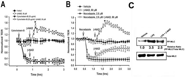
Effect of cytoskeletal alterations on β-NAD-induced endothelial cell barrier protection. (A) HPAEC were pretreated with either vehicle or cytochalasin B for 30 min and then stimulated with 50 μM β-NAD in TER measurement assay. Actin depolymerization decreased TER and completely prevented the effect of β-NAD on TER. (B) HPAEC were pretreated with either vehicle or the microtubule-disrupting agent, nocodazole, for 30 min and then stimulated with 50 μM β-NAD. Disruption of microtubules decreased TER, but failed to alter β-NAD-induced increase in TER. Results are presented as mean ± SE and derived from three independent experiments (*p < 0.05 compared with control). (C) β-NAD treatment decreases myosin light chain (MLC) phosphorylation stimulated by LPS. HPAEC treated by either vehicle or LPS alone, or LPS/β-NAD mixture for 4 hrs were lysed and analyzed by SDS-PAGE followed by immunoblotting with anti-phosphoMLC antibody. Immunoblotting with anti-MLC antibody was used as a loading control. Data obtained indicate that barrier-protective mechanism of β-NAD may be realized via stimulation of MLC phosphatase activity, decreasing phosphoMLC levels and preventing, therefore, actin stress fiber formation.

