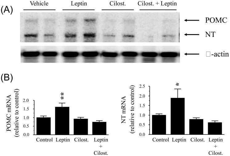
Fig. 1.
Proopiomelanocortin (POMC) and neurotensin (NT) gene expression as determined by ribonuclease protection assay in the hypothalamus following intra-cerebroventricular injection of leptin alone or in combination with cilostamide (Cilost.), a selective PDE3 inhibitor. (A): representative phosphorimages showing the level of POMC mRNA, NT mRNA and β-actin mRNA in the hypothalamus. (B): results obtained by phosphor imaging showing the changes in POMC and NT mRNA levels. The values were first normalized to β-actin mRNA levels and then expressed as relative to vehicle (artificial cerebrospinal fluid + dimethyl sulfoxide) control. Values represent the mean ± SEM. Control: n = 4, leptin: n = 5, cilost. : n = 5, and leptin + cilost. : n = 7. * p < 0.05 and ** p < 0.01 vs. all other groups.

