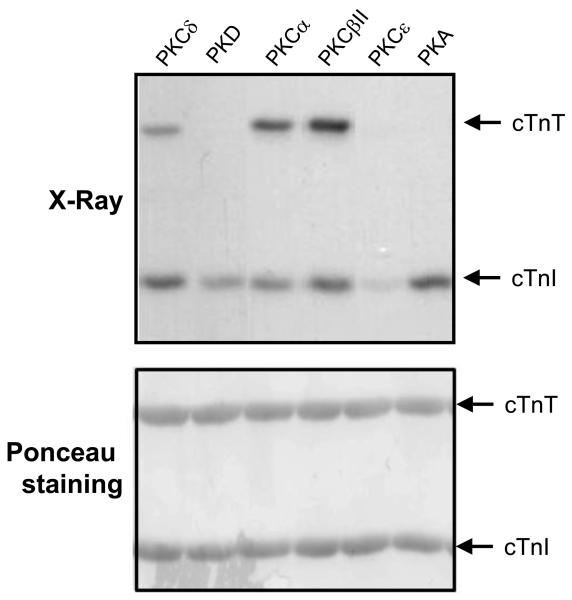
Figure 2. Differential phosphorylation of cTnT and cTnI by PKC-α, βII, δ, ε, PKD and PKA.
Recombinant troponin complexes were subjected to in vitro kinase assays (top) in buffers containing [γ-32P]ATP and either PKCα, PKCβII, PKCδ, PKCε, PKD or PKA. Phosphorylated forms of cTnT and cTnI were separated by SDS-PAGE and visualized by autoradiography. Ponceau staining (bottom) shows equal protein loading in each lane. Results were replicated in three separate experiments.

