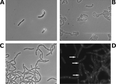
FIG. 2.
Microscopic observation of wild-type B. coagulans (A and B) and the ΔsigF strain (C and D). Bright spores are observed in wild-type cells using confocal microscopy during early (A) and late (B) stationary phase. No spores are observed in the ΔsigF strain (C), while asymmetric septa can be visualized using FM 9-95 membrane stain during fluorescence microscopy (D).

