
Figure 2. SKBR3-FoxM1 cell lines fail to accumulate p27 after treatment with Herceptin.
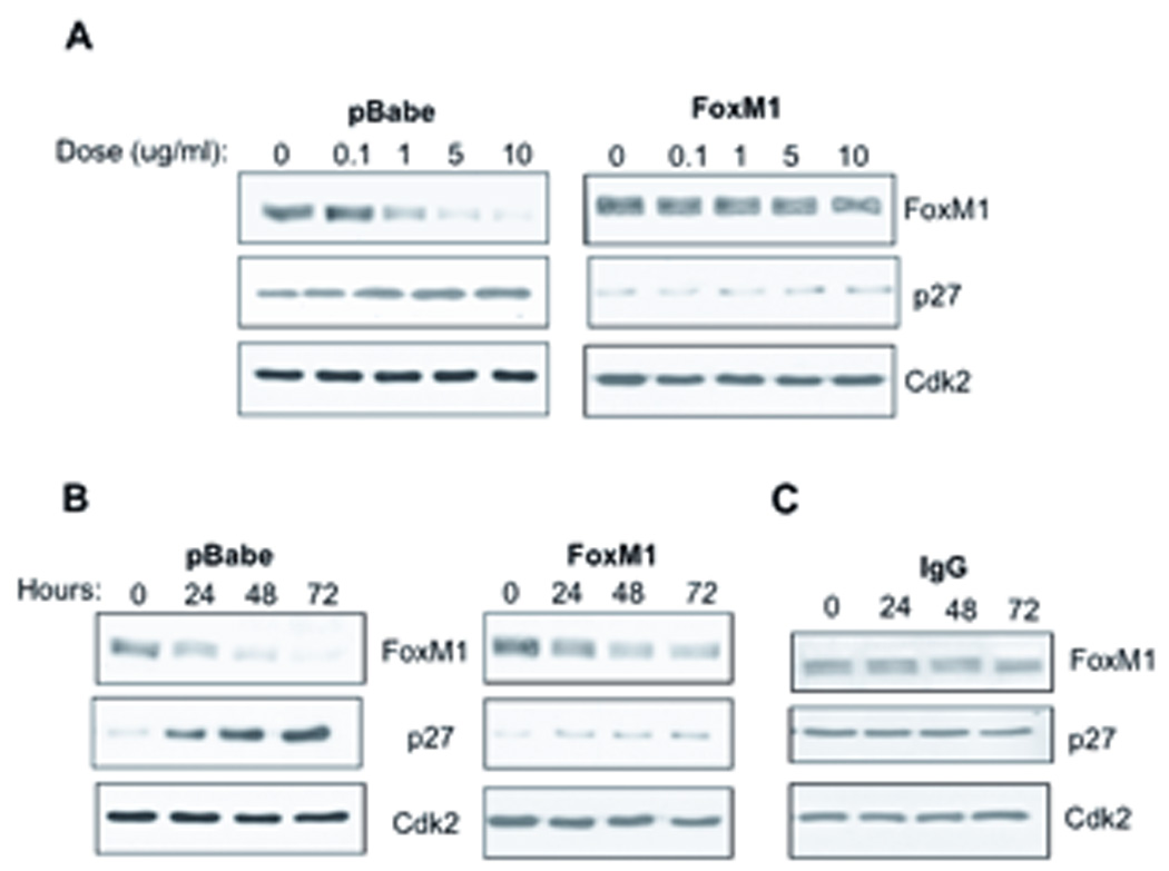
(A) SKBR3-pBabe and FoxM1 expressing cell lines were treated with increasing doses of Herceptin for 48 hours. Representative western blots of FoxM1 and p27 levels are shown. Cdk2 was used as a loading control. (B) SKBR3 stable cell lines were treated with 10ug/ml of Herceptin for 24, 48, and 72 hours. FoxM1 and p27 levels are shown by western blot. (C) SKBR3-pBabe cell lines were treated with 10ug/ml of IgG for indicated periods of time. FoxM1 and p27 were assayed by western blot and Cdk2 was used as a loading control.