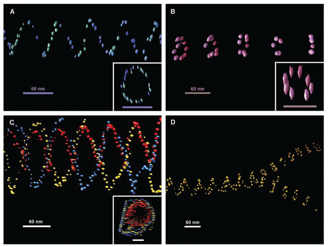
Fig. 3.
Representative 3D structures of nanoparticle tubes reconstructed from cryoelectron tomographic imaging. (A) One view of the tomogram of a single-spiral tube of 5-nm AuNPs. The inset shows a top view from the axis of two helical turns of the spiral tube; scale bar, 60 nm. (B) Tomogram of a stacked-ring tube of 5-nm particles. The inset shows a top view from the axis of a single ring from the stacked-ring tube; scale bar, 60 nm. (C) Tomogram of a double-spiral tube of 5-nm AuNPs with a single spiral of 5-nm nanoparticles inside each coded with a different color. The inset shows a top view from the axis of the double-wall spiral tube; scale bar, 60 nm. (D) Tomograph showing the splitting of a wider single-spiral tube into two narrower stacked-ring tubes of 10-nmAuNPs. All of the spiral tubes show a left-handed chirality. A weakly colored depth cue was applied to each view. The elongated appearance of the gold bead in the top views of the tubes is an effect of limited tilts in the tomography data collection. Movies of electron tomographic reconstruction corresponding to these structures are available in (28).

