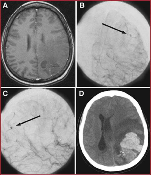
Figure 4.

Illustrative Case 1. Axial T1-weighted MRI with gadolinium (A), and anteroposterior (B) and lateral (C) projections of a cerebral angiogram after left internal carotid injection demonstrated minimal persistent AVM after radiosurgical treatment. Small early pooling of contrast is seen during the arterial phase (arrow) (B-C). A non-contrast head CT (D) demonstrated a large intraparenchymal hemorrhage with severe midline shift and transtentorial herniation.
