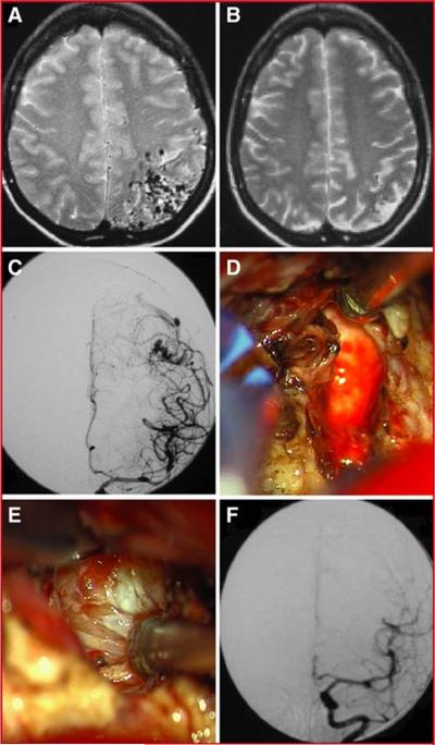
Figure 5.
Illustrative Case 2. Corresponding T2-weighted MRIs before (A) and after (B) radiosurgical treatment demonstrate a significant decrease in AVM size. An anteroposterior projection of a left internal carotid injection demonstrates the residual AVM after radiosurgical treatment (C). Intraoperative images demonstrate the gliotic margin (D) and sclerotic arterial feeders (E) that are frequently encountered after radiosurgical treatment. A post-operative angiogram demonstrates complete resection of the AVM (F).

