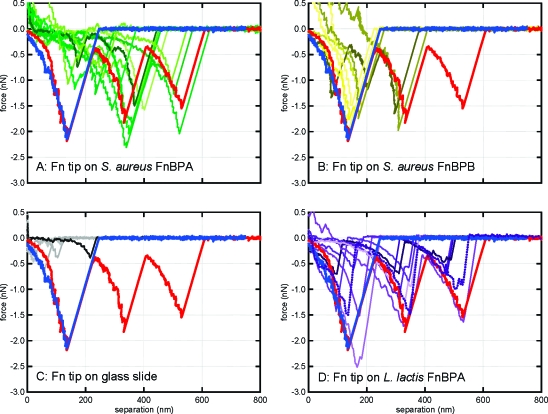
Figure 2.
Force spectra collected by atomic force microscopy in buffered saline. Shown are randomly selected retraction curves that exhibited a sawtooth-shaped binding event from a total of 4574 force profiles. A Fn-coated tip was used on (a) S. aureus that expresses only FnBPA (green); (b) S. aureus that expresses only FnBPB (yellow); (c) a glass slide (gray), and (d) recombinant L. lactis that expresses FnBPA or FnBPB (purple). The blue and red curves on each panel are taken directly from previous work with clinical isolates as reported in ref (7). The blue and red spectra are from nasal carriage isolates of S. aureus and invasive isolates of S. aureus, respectively.(7)

