
Figure 2.
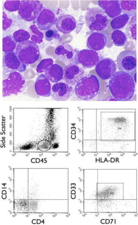
Post-UCBT diagnosis of AML. A Wright-stained bone marrow aspirate smear (×1000) showing blasts with histological characteristics consistent with acute monoblastic leukemia, including a moderate amount of finely granular cytoplasm with the absence of Auer rods, lacy chromatin with one to several prominent nucleoli, and occasional nuclear indentations or grooves (top). Flow cytometric analysis reveals a blast population with a CD45 expression level below normal lymphocytes and a low side scatter (circled/gated cells, top left). Analysis of these cells reveals strong expression of the early hematopoietic associated antigens CD34 and HLA-DR (top right), partial dim expression of the monocytic marker CD4 and lack of expression of the monocytic marker CD14 (bottom left), and dim expression of the myeloid marker CD33 and moderate expression of CD71, which is present on proliferating cells (bottom right). This immunophenotypic profile is consistent with acute monoblastic leukemia.