Figure 2.
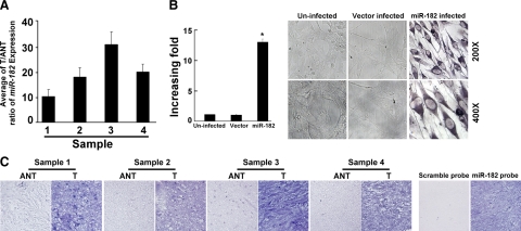
Expression of miR-182 is elevated in primary gliomas as compared with tumor-adjacent tissues of the same individuals with gliomas. A: Real-time RT-PCR analyses of miR-182 expression in paired primary glioma tissues (T) and glioma ANT of four individual patients. Expression levels were normalized for U6, and the average ratio of miR-182 expression was quantified. B: Ectopic expression of miR-182 in NHA cells analyzed by real-time RT-PCR (left) and ISH (right). Statistical significance of *P < 0.05. C: miR-182 expression level up-regulated in the paired primary T and glioma ANT examined by ISH. Validation for the specificity of the in situ probe of miR-182 is shown. Sister sections of an anaplastic astrocytoma, World Health Organization grade III were hybridized with the miR-182 probe or a scrambled control probe. Error bars (SD) were calculated from triplicate samples.

