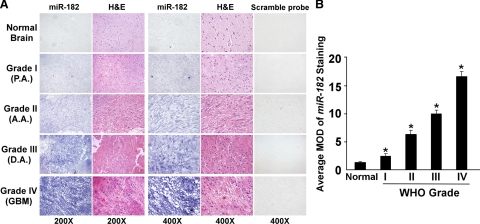
Figure 3.
miR-182 expression correlates with glioma progression. A: Representative images of ISH analyses of a total of three normal brain tissues and 253 primary glioma specimens, including World Health Organization (WHO) grade I to IV tumors, were stained by ISH by using an miR-182 probe. Sister sections were also stained with a scrambled control probe. ISH analyses were performed two independent times on sections of each specimen with similar results. B: statistical analyses of the average MOD of miR-182 staining between normal brain tissues (three cases) and glioma specimens of different World Health Organization grades (29 random cases per grade). Average MOD of miR-182 staining increases as gliomas progress to higher grades. Statistical significance of *P < 0.05.

