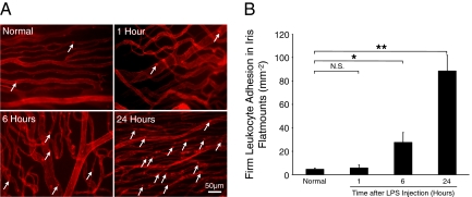
Figure 10.
Time course of leukocytes accumulation in the iris vasculature. To quantify firm leukocyte adhesion, normal and LPS-injected animals were perfused with rhodamine ConA and iridal flat mounts were prepared. A: Representative micrographs indicate ex vivo visualization of firmly adhering leukocytes in the iridal microvasculature of normal or LPS-treated animals at different time points. Arrows indicate firmly adhering leukocytes (bright red spots) inside the iridal microvessels. B: Quantitative comparison of the numbers of accumulated leukocytes in iridal microvasculature per surface area (mm−2), with or without LPS injection. *P < 0.05; **P < 0.01; N.S., not significant.

