Figure 3.
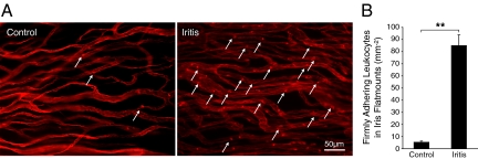
Conjugated MSs detect firmly adhering leukocytes. Twenty-four hours after LPS treatment, Lewis rats were perfused with PBS and rhodamine ConA to stain the vasculature and adherent leukocytes. Iris flat mounts of EIU animals were prepared. A: Ex vivo visualization of firmly adhering leukocytes in the iris microvasculature of normal and iritic animal. Arrows delineate firmly adhering leukocytes (bright red spots) inside the iridal microvessels. B: The number of firmly adhering leukocytes in iridal microvasculature per iris surface area (mm−2) in control and EIU animals. **P < 0.01.

