Figure 4.
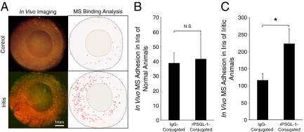
Noninvasive in vivo imaging of MS adhesion in iridal microvasculature. Anesthetized normal and iritic rats were systemically injected with rPSGL-1-conjugated fluorescent MSs, the interaction of which in the iris vasculature was visualized by intravital microscopy. A: Video micrographs of rPSGL-1-conjugated MS accumulation in vivo, 30 minutes after injection. In the video still images (left), yellow-green spots delineate adhering MSs. In the Image J pictures (right), each dot indicates one automatically counted adhering MS in the iris vessels of normal and iritic animals. B: Quantification of the in vivo accumulation of IgG- and rPSGL-1-conjugated MSs in the iris vasculature of normal animals. N.S., not significant. C: Quantification of the in vivo MS accumulation IgG- and rPSGL-1-conjugated MSs in iris vessels of uveitic animals. *P < 0.01.

