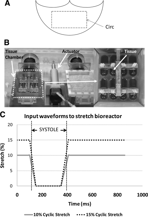
Figure 1.
Preparation of aortic valve cusps for the experiment. A circumferentially oriented 15 × 10 mm section was excised from the central region of porcine aortic valve cusps (A). An ex vivo tensile stretch bioreactor was used in this study (B).22 A magnified image of the tissue chamber is shown on the right. A loading curve was used in this study. The gradients of the extension and relaxation approximated those experienced in vivo (C).20

