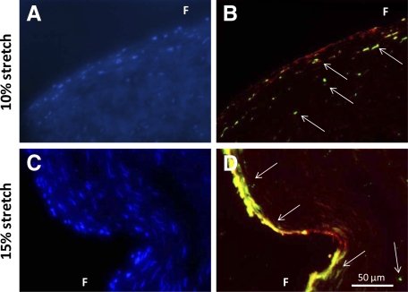
Figure 7.
Representative micrographs of BMP-2 (green) and von Willebrand Factor (red) double immunohistochemical staining for samples stretched at 10% (A and B) and 15% (C and D) in fully osteogenic media. Cell nuclei are stained with 4′,6-diamidino-2-phenylindole (blue) and presented in a separate image for clarity of viewing. Arrows represent immunopositive cells. F indicates fibrosa.

