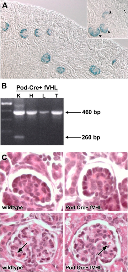
Figure 1.
The Podocin-Cre strain recombines floxed alleles in podocytes of the kidney. A: Pod-Cre mice were mated to the floxed LacZ (β-galactosidase) reporter mouse R26R, and progeny were genotyped for Cre. Both Cre+ and Cre− kidneys were developed for β-galactosidase activity. Blue, β-galactosidase positive cells are confined to podocytes of developing glomeruli, beginning at the early capillary loop stage (inset, arrowheads), exclusively in Cre+ kidneys (n = 5). No β-galactosidase positive cells were seen in Cre− kidneys (n = 4), or in late S-shaped developing glomeruli (inset, arrow). B: Pod-Cre mice were crossed to fVHL mice generating a podocyte-specific deletion of VHL. Genomic DNA was isolated from Pod-Cre fVHL tissues (K = kidney, H = heart, L = liver, and T = tail). Primers flanking the floxed VHL locus amplified the nonrecombined VHL locus (460 bp) in all tissues. The smaller, recombined product (260 bp) is only seen in kidney tissue. C: Glomerular development is normal in Pod-Cre fVHL mice. Pod-Cre × fVHL litters were screened for amplification of Cre and presence of the floxed VHL locus. Animals that were either homozygous wild-type at the VHL locus, or were Cre-negative, served as wild-type littermate controls (wild-type, left). Pod-Cre fVHL mice were both Cre-positive and homozygous for the fVHL locus (Pod-Cre fVHL). At postnatal day five, kidney morphology of H&E paraffin sections shows normal capillary loop stage glomeruli in both wild-type and Pod-Cre fVHL (top). Mature glomeruli also appear normal in both wild-type and Pod-Cre fVHL animals (bottom) with red blood cells present in capillary loops (arrows).

