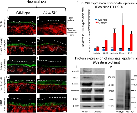
Figure 2.
Reduced epidermal differentiation-associated molecules and defective conversion of profilaggrin to filaggrin in Abca12−/− neonates. A and B: Immunofluorescence staining for kallikrein 5 (KLK5) (Alexa488, green), one of the lamellar granule (LG) contents, was weak in the Abca12−/− neonatal mice granular keratinocyte layers (B), in contrast to its intense labeling in the granular layers and lower cornified layers of wild-type neonatal skin (A). C and D: In situ transglutaminase 1 (TGase1) activity assay (fluorescein isothiocyanate, green) with dansyl-cadaverine showed the granular layer keratinocytes in the Abca12−/− neonates had weak transglutaminase 1 activity only in the cytoplasm (D), compared with distinct transglutaminase 1 activity with the peripheral pattern throughout granular layer keratinocytes in the wild-type neonates (C). Cytoplasmic localization of transglutaminase 1 in Abca12−/− neonates indicated its inability to bind to the cell membrane and to therefore function at its proper place despite the significantly enhanced mRNA expression of transglutaminase 1 (see Figure 2K). E and F: Immunofluorescence staining showed loricrin (Alexa 488, green) expressed sparsely in the Abca12−/− neonatal mice granular layer (F), compared with its intense expression in the granular layer of the wild-type neonatal skin (E). G and H: Both Abca12−/− and wild-type neonatal skin showed intense profilaggrin/filaggrin (proFLG/FLG) (Alexa488, green) expression in the granular layer keratinocytes. However, in Abca12−/− neonatal skin, profilaggrin/filaggrin distribution was also observed throughout the cornified layers. I and J: Desmoglein 1(DSG1) (Alexa 488, green), a cell adhesion molecule unassociated with keratinization, was expressed at the cell periphery in the lower granular and spinous layers of the both Abca12−/− neonatal skin and wild-type mouse skin. (nuclear stain; propidium iodide, red, dotted lines, the skin surface). Original magnification ×40; Scale bars, 20 μm. K: mRNA expression of loricrin, kallikrein 5 (KLK5), involucrin, transglutaminase 1 (TGase1) and filaggrin (FLG) was up-regulated in the Abca12−/− neonatal epidermis. (Abca12−/− neonates, n = 5; wild-type neonates, n = 5, mRNA expression levels of wild-type neonatal epidermis = 1). L: Western blotting of epidermal extracts showed that protein expression of kallikrein 5 (KLK5) and loricrin was lower in the Abca12−/− neonatal epidermis (right) than in the wild-type neonatal epidermis (left). There were no significant differences of desmoglein 1(DSG1), involucrin, β-actin expressions between the Abca12−/− and wild-type epidermis. M: Western blotting with anti-profilaggrin/filaggrin antibody revealed the Abca12−/− neonatal epidermis (right) expressed more profilaggrin/filaggrin protein than wild-type neonatal epidermis (left). High molecular weight smear band corresponding to non-converted profilaggrin peptides were characteristic to the Abca12−/− neonatal epidermis. Western blotting using serial protein dilutions is shown in the supplemental Figure 2 (see Supplemental Figure S2 at http://ajp.amjpathol.org). KLK5, kallikrein 5; LG, lamellar granule; TGase1, transglutaminase 1; CCE, cornified cell envelope; FLG, filaggrin; KHG, keratohyalin granule; DSG1, desmoglein 1; proFLG, profilaggrin; 4FLG, filaggrin tetramer; 3FLG, filaggrin trimer; 2FLG, filaggrin dimer; 1FLG, filaggrin monomer.

