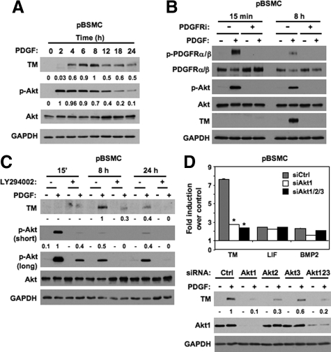
Figure 2.
Expression of TM in pBSMC is regulated by a PDGFR/Akt pathway. A: Lysates of quiescent pBSMC stimulated with PDGF for the indicated times were resolved by SDS-PAGE and immunoblotted with the indicated antibodies. The values represent quantification of PDGF-induced TM levels and of pAkt levels normalized to total Akt at the indicated times. In each case the maximum signal was set to a value of one, and the intensity of other signals was determined relative to this reference. B: pBSMC pretreated with vehicle (dimethyl sulfoxide) or 10 μmol/L PDGFR inhibitor (PDGFRi) for 30 minutes were stimulated without (−) or with (+) 30 ng/ml PDGF for the indicated times. Lysates were resolved by SDS-PAGE and immunoblotted with the indicated antibodies. Rapid activation (phosphorylation) of the PDGFR and Akt was observed at 15 minutes, with activation attenuated by eight hours. TM protein expression was detected at 8 hours. The PDGF-induced increase in TM levels, as well as PDGFR and Akt activation, was abolished by PDGFR inhibition but not with vehicle alone (dimethyl sulfoxide). C: Lysates of pBSMC treated without (−) or with (+) 30 ng/ml PDGF for the indicated times following 30 minutes pretreatment with vehicle (dimethyl sulfoxide) or 10 μmol/L LY294002, were resolved by SDS-PAGE and immunoblotted with the indicated antibodies. PDGF-induced phosphorylation of Akt and induction of TM protein expression was attenuated at all time-points by LY294002, whereas total Akt levels were unchanged. The values represent quantification of PDGF-induced TM levels at eight and 24 hours, and of pAkt levels (normalized to total Akt) at 15 minutes, 8 hours, and 24 hours in the absence and presence of LY294002. As for A, the maximum signal was set to a value of 1, and the intensity of other signals was determined relative to this reference. A long exposure for phospho-Akt is included to illustrate presence of signal at the 8- and 24-hour time points in cells treated with PDGF, consistent with the data in Figure 2, A and B. D: Upper panel, Total RNA isolated from pBSMC nucleofected with nontargeting siRNA duplexes or duplexes against Akt1, Akt2, Akt3, or all three Akt isoforms together, and treated without or with PDGF for eight hours was reverse transcribed to cDNA and amplified with primers for TM, LIF, BMP2, or glyceraldehyde-3-phosphate dehydrogenase. Data are presented as mean fold change in gene expression with PDGF treatment compared with nontreated controls. Only the reduction in TM expression was statistically significant with Akt knockdown (*P < 0.01 relative to siCtrl), with LIF and BMP2 showing little or no change with Akt silencing. Lower panel: Lysates from pBSMC nucleofected with siRNAs to Akt1, Akt2, Akt3 individually or combined, or nontargeting control siRNA were resolved by SDS-PAGE and immunoblotted with the indicated antibodies. The values represent quantification of the change in PDGF-induced TM levels following Akt isoform knockdown.

