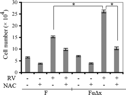
Figure 4.
NAC blunted vitreous-driven proliferation. Cells were plated at a subconfluent density in serum-free medium that was supplemented with nothing, vitreous, or vitreous and NAC (10 mmol/L). Vitreous from healthy rabbits was diluted 1:2 in DMEM, and 0.5 ml of the resulting solution was added to the appropriate wells of a 24-well plate. The medium was replaced every day, and cells were counted on day three with a hemocytometer. The data shown are the mean ± SD from three independent experiments; there is a statistically significant difference, *P < 0.05. The data indicate that NAC blunted vitreous-driven proliferation of both cell lines and that the PDGFRα-expressing cells lost their proliferative advantage in the presence of NAC.

