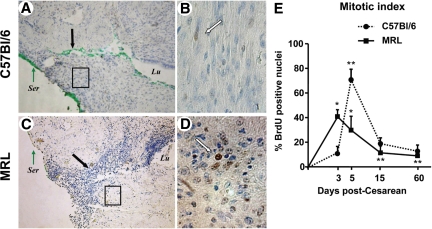
Figure 4.
BrdUrd nuclear labeling of the uterine scar 3 days post-Cesarean delivery in C57Bl/6 and MRL/MpJ+/+ (MRL) mice. BrdUrd labeled nuclei (brown) could be identified into the wound site of both C57Bl/6 (A) and MRL (C) mice. Original magnification, ×100. The green dye marks the site of uterine wound at the time of tissue collection and is indicated by the black arrows. B and D show the respective boxed areas at higher magnification (×400). White arrows mark BrdUrd labeled nuclei. E: Mitotic index (ratio of BrdUrd labeled nuclei-to-total counted nuclei). Statistical analysis: two-way analysis of variance followed by posthoc Student-Newman-Keuls tests: **P < 0.05 versus day three of same strain. *P < 0.05 versus C57Bl/6 phenotype. Ser, uterine serosa; Lu, uterine lumen.

