Figure 5.
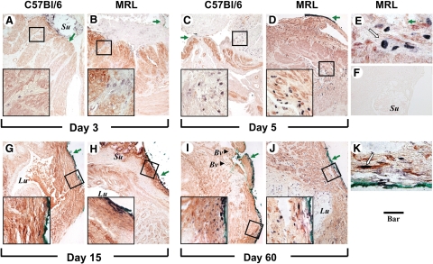
Topographical co-localization of BrdUrd and smooth muscle actin post-Cesarean delivery in C57Bl/6 and MRL/MpJ+/+ (MRL) mice. Cells with BrdUrd labeled nuclei (purple black) could be identified between the red muscle bundles in C57Bl/6 [day 3 (A) and day 5 (C)] and MRL [MRL: day 3 (B) and day five (D)] mice. At the later times, cells positive for both BrdUrd and smooth muscle actin could be observed in proximity to serosa in the scar area both in C57Bl/6 [day 15 (G) and day 60 (I)] and MRL [MRL: day 15 (H) and day 60 (J)] mice. Original magnification, ×100 (Scale bar = 100 μm). The areas delineated by the squares are further shown in the left lower corner of each panel as higher magnification captions of the boxed areas (×640; Scale bar = 33 μm). E and K show representative areas at ×1000 magnification of D (day five) and J (day 60) in MRL mice. F shows a section from an MRL animal (day five) processed identically but omitting the two primary antibodies (Scale bar = 100 μm). The green arrows indicate the site of the uterine scar as marked with tissue dye (green or black). The white arrow in K points to a BrdUrd+/α-SMA+ cell in the subserosal area of regenerated uterine wall. Su, suture material; Lu, uterine lumen; Bv, blood vessel (arrowhead).

